
PEPTIDES PARTNERS BLOG · November 11, 2026
The Research Science of KPV: Cellular Repair Mechanisms, Lineage, and Laboratory Sourcing
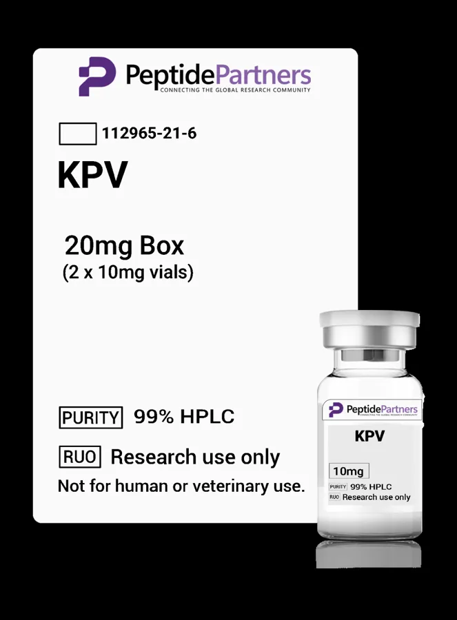
The tripeptide KPV (consisting of the amino acid sequence Lysine-Proline-Valine) has emerged as a focused subject of interest within cellular repair, epithelial biology, and inflammatory signaling research. Derived from the C-terminal end of alpha-melanocyte-stimulating hormone (α-MSH), KPV encompasses residues 11 through 13 of the parent neuropeptide. Despite its small molecular footprint—possessing a molecular weight of approximately 383.49 g/mol—the molecule has been extensively investigated for its ability to replicate many of the protective and signaling attributes of α-MSH without triggering melanogenesis. This educational overview outlines the origin of KPV, its mechanistic pathways observed in preclinical models, and standard evaluation criteria for researchers sourcing this peptide from laboratory-grade catalogs such as Peptides Partners.
Historical Lineage: From Neuropeptide to Targeted Tripeptide
Research into KPV originated from broader neuroendocrinology studies focusing on the melanocortin system. Alpha-MSH, a 13-amino-acid peptide cleaved from proopiomelanocortin (POMC), was historically recognized for its influence on skin pigmentation through Melanocortin 1 Receptor (MC1R) agonism. However, during the late 20th century, investigators noted that systemic and local delivery of α-MSH exerted potent regulatory effects on localized cellular inflammation and tissue integrity, even in models lacking functional pigmentary responses.
To determine the minimal active sequence responsible for these cytoprotective actions, researchers conducted structure-activity relationship (SAR) experiments. These investigations systematically truncated the α-MSH sequence and revealed that the terminal tripeptide, Lys-Pro-Val, retained significant anti-inflammatory and cellular repair signaling capacities. Crucially, KPV demonstrated an ability to operate independently of classical melanocortin receptor activation, allowing scientists to study cellular preservation pathways isolated from pigment-inducing neuroendocrine cascades.
Mechanisms Investigated in Preclinical Research
In vitro assays and animal models have explored KPV across diverse tissue types, with particular concentration on mucosal surfaces, dermal tissue, and gastrointestinal epithelia. The literature highlights several core biochemical and cellular mechanisms.
Intracellular Transport via PepT1
Unlike larger polypeptides that depend on receptor-mediated endocytosis or specialized pinocytosis, KPV has been shown to enter target cells via solute carrier family 15 member 1 (SLC15A1), commonly referred to as the peptide transporter 1 (PepT1). This transport mechanism is especially prevalent in epithelial cell types, such as those lining the intestinal tract. Preclinical models indicate that upregulation of PepT1 during cellular stress facilitates direct cytoplasmic entry of KPV, enabling rapid intracellular signaling without requiring high-affinity cell-surface receptor binding.
Modulation of the NF-κB Signaling Cascade
Once inside the cytoplasm, KPV has been evaluated for its ability to regulate the Nuclear Factor kappa B (NF-κB) transcription factor network. Under typical inflammatory stress, the phosphorylation and subsequent degradation of the inhibitory protein IκBα allows NF-κB to translocate to the nucleus, where it triggers the transcription of pro-inflammatory cytokines, chemokines, and matrix metalloproteinases (MMPs). In cell culture models, researchers have observed that KPV inhibits the translocation of NF-κB p65 subunits to the cell nucleus, leading to a measurable reduction in down-stream inflammatory mediators such as IL-1β, IL-6, and TNF-α.
Support of Epithelial Barrier Integrity
In cellular repair assays, investigators frequently employ cell-line monolayers (such as Caco-2 or epidermal keratinocytes) subjected to chemical or mechanical disruption. In these controlled settings, KPV exposure has been associated with the preservation of tight junction proteins, including zonula occludens-1 (ZO-1) and occludin. By maintaining junctional complex architecture, the tripeptide has demonstrated capacity in laboratory literature to reduce paracellular permeability and preserve cellular polarity during environmental challenges.
Comparative Overview of KPV Preclinical Research Targets
The following table summarizes the primary research targets and biological systems detailed in published preclinical literature involving KPV:
| Research Target | Primary Model Systems | Key Observed Interactions |
|---|---|---|
| NF-κB Pathway | Epithelial and immune cell lines (e.g., Caco-2, macrophages) | Inhibition of nuclear p65 translocation; reduction in pro-inflammatory cytokine expression. |
| PepT1 Transporter Kinetics | Intestinal brush border membrane vesicles, cell assays | Facilitated entry of tripeptide via proton-coupled oligopeptide transport mechanisms. |
| Epithelial Barrier Dynamics | Dermal keratinocyte cultures, murine mucosal models | Maintenance of junctional scaffolding (ZO-1, occludin); modulation of matrix turnover. |
| Microbial Dynamics | Direct bacterial/fungal pathogen cultures (e.g., C. albicans, S. aureus) | Evaluation of non-lytic growth inhibition and structural membrane modulation. |
Evaluating Research-Grade KPV: Documentation and Analytical Standards
When procuring KPV for strictly laboratory or in vitro research purposes, consistency in the molecular identity and synthesis profile is essential to maintain experimental reproducibility. Investigators assessing peptide suppliers should focus on specific analytical parameters and packaging methodologies.
High-Performance Liquid Chromatography (HPLC) and Mass Spectrometry (MS)
Authentic research documentation requires rigorous analytical testing for every synthesis lot. High-Performance Liquid Chromatography (HPLC) provides an empirical measure of purity, isolating the desired KPV sequence from truncated or deletion sequences that may occur during solid-phase peptide synthesis. Mass Spectrometry (typically LC-MS or MALDI-TOF) confirms the exact molecular weight and elemental composition, ensuring no synthesis adducts or unintended modifications have occurred.
Net Peptide Content and Counter-Ion Analysis
Standard lyophilized peptides contain residual moisture, counter-ions (such as trifluoroacetate [TFA] or acetate), and the peptide itself. Standard practice involves distinguishing between gross lyophilized weight and net peptide content. Researchers investigating sensitive cellular signaling pathways often monitor residual counter-ion concentrations, as elevated TFA levels can alter baseline cellular viability in cell-culture assays.
Multi-Vial Packaging versus Single Vials
Peptide degradation pathways—primarily hydrolysis and oxidation—are accelerated by repeated freeze-thaw cycles. In laboratory environments conducting longitudinal assays, utilizing multi-vial packs (e.g., 5-vial or 10-vial formats) is generally preferred over single large-quantity vials. Sourcing smaller aliquots minimizes exposure to atmospheric moisture and temperature fluctuations once reconstituted for in vitro protocols.
Sourcing KPV from Peptides Partners
Peptides Partners provides research-use-only peptides synthesized to precise laboratory specifications. Within the cellular repair catalog, KPV is offered in defined vial packs engineered for research workflows requiring analytical traceability and batch-to-batch consistency. Each batch is accompanied by comprehensive analytical documentation, allowing research personnel to verify identity and purity profiles prior to experimental integration. All materials available through the catalog are intended strictly for laboratory investigation and in vitro analysis.
Where to buy KPV for research
Peptides Partners lists KPV (10mg vials) in 3 pack sizes, all in stock, with price per mg shown for each option. Browse more cellular repair compounds in the Peptides Partners research peptide shop. All products are for laboratory research use only.