
PEPTIDES PARTNERS BLOG · November 11, 2026
The Research Science of Humanin: Investigating the First Mitochondria-Derived Peptide
Mitochondria have historically been categorized primarily as metabolic hubs responsible for generating cellular adenosine triphosphate (ATP) through oxidative phosphorylation. Over the past two decades, however, research has illuminated a secondary role for these organelles: retrograde signaling via retrograde communications mediated by small, biologically active peptides encoded directly within mitochondrial DNA (mtDNA). The prototypical member of this class of signaling molecules is Humanin, a 24-amino-acid peptide that sparked widespread scientific interest in non-canonical mitochondrial genetics and cell-survival pathways. Within laboratory investigations, Humanin remains a prominent subject across neurobiology, cellular senescence, and metabolic biochemistry.
Discovery and Genetic Origins of Humanin
Humanin was first identified in 2001 by a research group led by Dr. Ikuo Nishimoto at Keio University. Using functional expression screening of cDNA libraries derived from the unaffected occipital cortex of post-mortem Alzheimer’s disease brain tissue, researchers isolated a novel sequence that rescued neuronal cells from death induced by various familial Alzheimer’s disease genes and amyloid-beta toxicity. Subsequent genomic mapping revealed that this open reading frame was not situated within the nuclear genome, but rather resided inside the 16S ribosomal RNA (MT-RNR2) region of the circular mitochondrial chromosome.
This discovery disrupted conventional molecular dogma, which held that human mtDNA strictly encoded 13 respiratory chain subunits, 22 transfer RNAs (tRNAs), and 2 ribosomal RNAs (rRNAs). Humanin demonstrated that mitochondria also transcribe short open reading frames (sORFs) capable of producing functional, translatable peptides. In humans, the canonical sequence consists of 24 amino acids (Met-Ala-Pro-Arg-Gly-Phe-Ser-Cys-Leu-Leu-Leu-Leu-Thr-Ser-Glu-Ile-Asp-Leu-Pro-Val-Lys-Arg-Arg-Ala). Research has demonstrated that endogenous Humanin can act both cell-autonomously as an intracellular factor and non-cell-autonomously as a secreted endocrine or paracrine messenger.
Primary Molecular Mechanisms Investigated in Research
Investigators studying Humanin focus on distinct, multi-modal biochemical pathways, categorizing its biological actions into intracellular protein-protein interactions and cell-surface receptor engagements.
Intracellular Interaction with BAX
One of the earliest documented intracellular mechanisms of Humanin is its direct interaction with BAX (Bcl-2-associated X protein), a core pro-apoptotic regulator. In baseline cellular conditions, BAX resides largely in the cytosol. Upon cellular stress, BAX undergoes a conformational shift and translocates to the outer mitochondrial membrane, facilitating membrane permeabilization and the release of cytochrome c. In vitro binding assays have shown that intracellular Humanin binds cytosolic BAX, physically sequestering it and impeding its membrane translocation, thereby preventing caspase activation and downstream apoptotic cascades.
Extracellular Receptor Complex Engagement
When acting extracellularly, synthetic and native Humanin peptides have been documented to bind two distinct receptor configurations:
- The Trimeric Interleukin Receptor Complex: Humanin engages a heterotrimeric receptor assembly composed of the common signaling subunit glycoprotein 130 (gp130), the WSX-1 subunit (IL-27R alpha), and the ciliary neurotrophic factor receptor alpha (CNTFR-alpha). Binding to this complex initiates the phosphorylation of Janus kinase 2 (JAK2) and downstream Signal Transducer and Activator of Transcription 3 (STAT3), as well as extracellular signal-regulated kinase (ERK) pathways.
- Formyl Peptide Receptor-Like 1 (FPRL1 / FPR2): Humanin also functions as a ligand for the G-protein coupled receptor FPRL1 (also known as formyl peptide receptor 2). Binding triggers intracellular calcium mobilization and activates survival kinases, including protein kinase B (Akt).
Cross-Talk with Metabolic Regulators
Beyond apoptosis, Humanin interacts with circulating proteins, notably Insulin-like Growth Factor Binding Protein 3 (IGFBP-3). Preclinical assays have identified that Humanin can physically bind IGFBP-3, altering its interaction with cell surface domains and modulating insulin-like growth factor (IGF) signaling dynamics. This interaction has made Humanin a molecule of interest in models of cellular metabolism, insulin resistance, and endocrine signaling.
Core Domains of Laboratory Investigation
Because Humanin functions as a general stress-response peptide, investigators utilize it across several distinct scientific fields to assess how cells mitigate environmental and biochemical stressors.
Neurodegenerative Disease Models: Preclinical research frequently assesses Humanin in cellular assays exposed to amyloid-beta oligomers, excitotoxic glutamate levels, and prion-derived peptide fragments. Researchers monitor cell viability, reactive oxygen species (ROS) accumulation, and mitochondrial membrane integrity.
Vascular and Endothelial Biology: Endothelial dysfunction driven by oxidized low-density lipoproteins (oxLDL) or inflammatory cytokines is a primary focus of cardiovascular studies. Preclinical research evaluates whether Humanin modulates endothelial nitric oxide synthase (eNOS) phosphorylation and cellular apoptosis in human umbilical vein endothelial cells (HUVECs).
Aging and Mitohormesis: Humanin concentrations have been observed to vary as a function of chronological age across various species. Consequently, geroscience laboratories explore Humanin within models of cellular senescence, examining its role in mitochondrial proteostasis, autophagy induction, and organismal resilience against oxidative stress.
Comparison of Research Models Studying Humanin
| Research Category | Primary Biological Model | Primary Molecular Endpoints |
|---|---|---|
| Neuronal Cytoprotection | Cortical neurons, PC12 cell lines | BAX translocation, Caspase-3 cleavage, Cell viability assays |
| Metabolic Signaling | Primary hepatocytes, 3T3-L1 adipocytes | STAT3 phosphorylation, IGFBP-3 binding affinity, Glucose uptake |
| Vascular Integrity | Endothelial cultures (e.g., HUVECs) | eNOS activation, ROS generation, Inflammatory cytokine release |
| Cellular Senescence | Dermal fibroblasts, Rodent tissue panels | Mitochondrial membrane potential, Senescence-associated beta-gal |
Sourcing Humanin: Evaluating Documentation and Formats
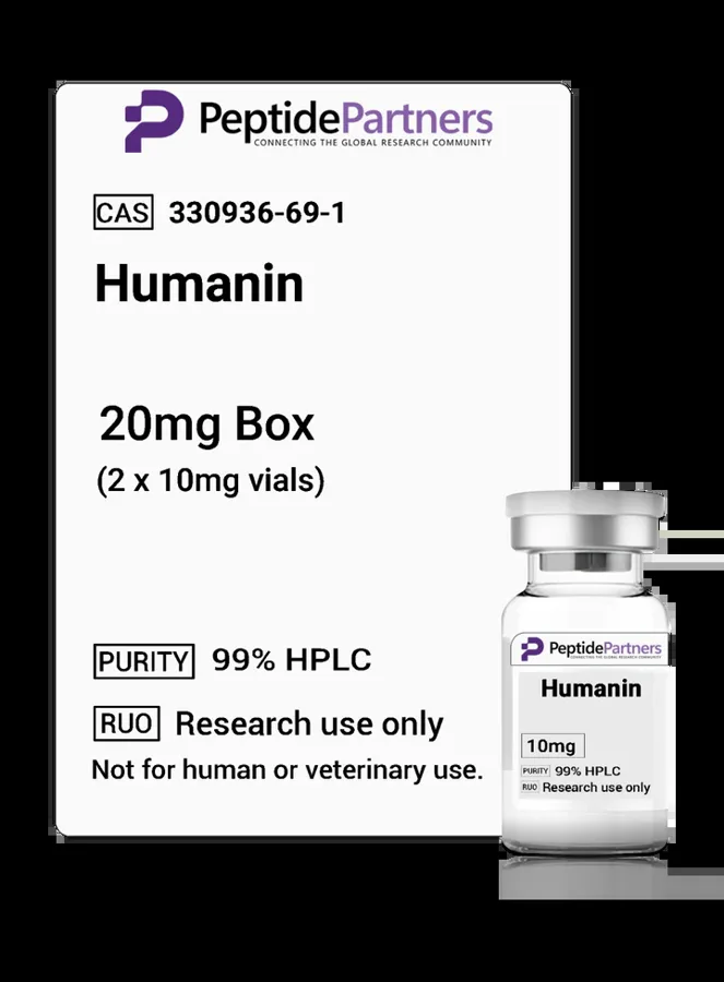
When securing Humanin from the Peptides Partners catalog, research facilities and independent laboratories evaluate multiple biochemical parameters to ensure experimental reproducibility. Because Humanin is a relatively hydrophobic, 24-amino-acid sequence with multiple leucine residues, peptide integrity, purity, and formulation require thorough analytical confirmation.
Analytical Documentation
Standard laboratory verification protocols require reviewing two primary analytical instruments prior to integrating a peptide into an experimental design:
- High-Performance Liquid Chromatography (HPLC): HPLC chromatograms confirm chemical purity by demonstrating a single, distinct peak under specified retention times, ensuring the absence of truncated synthesis products or peptide impurities.
- Mass Spectrometry (MS): Electrospray ionization or MALDI-TOF mass spectrometry confirms that the observed molecular weight corresponds precisely to the theoretical mass calculated for the peptide sequence.
Lyophilized Vial Packs and Experimental Design
Peptide stability is sensitive to repeated freeze-thaw cycles once reconstituted in sterile aqueous or buffered solutions. For this reason, laboratories often compare single-vial configurations against multi-vial packs when planning experimental protocols. Sourcing smaller-mass multi-vial packs allows researchers to reconstitute only the amount necessary for a given assay run, reducing peptide degradation over time and maintaining baseline consistency across long-term, multi-plate studies.
All materials supplied under this category are strictly designated for laboratory research, in vitro studies, and scientific investigation only. They are not intended for diagnostic, clinical, or therapeutic applications.
Where to buy Humanin for research
Peptides Partners lists Humanin (10mg vials) in 3 pack sizes, all in stock, with price per mg shown for each option. Browse more all products compounds in the Peptides Partners research peptide shop. All products are for laboratory research use only.