
PEPTIDES PARTNERS BLOG · November 11, 2026
The Research Science of Vasoactive Intestinal Peptide (VIP): Discovery, Mechanisms, and Laboratory Sourcing
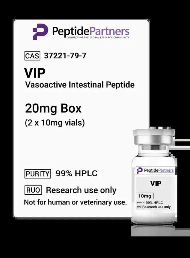
Vasoactive Intestinal Peptide (VIP) is an endogenous 28-amino-acid neuropeptide that plays a multifaceted role in biological signaling. Classified structurally within the secretin-glucagon superfamily, VIP operates across both the central and peripheral nervous systems, as well as the gastrointestinal, respiratory, and cardiovascular networks. In scientific literature, it is widely investigated as a potent vasodilator, a non-adrenergic non-cholinergic (NANC) neurotransmitter, and a modulator of cellular immune responses. For biochemical and biomedical researchers, VIP serves as a foundational tool for elucidating G-protein-coupled receptor (GPCR) cascades, neuro-immune cross-talk, and the physiological regulation of vascular smooth muscle tone.
Discovery and Evolutionary Context
VIP was initially isolated in the late 1960s and early 1970s by researchers Sami Said and Viktor Mutt from porcine small intestine extracts. Their early work identified a fraction capable of inducing sustained systemic vasodilation, an observation that inspired the peptide’s name. Subsequent investigations revealed that while the peptide was concentrated heavily in intestinal tissue, its distribution was far broader than initially understood.
From an evolutionary perspective, VIP exhibits remarkable sequence conservation among vertebrate species. The human sequence shares near-complete homology with porcine, bovine, and murine counterparts, differing by only a minimal number of conserved amino acid substitutions across divergent taxa. This evolutionary preservation underscores the essential nature of VIP-mediated signaling pathways in coordinating basic autonomic functions, tissue perfusion, and homeostatic cellular communication.
Molecular Structure and Receptor Dynamics
The VIP monomer consists of 28 amino acids folded into a single alpha-helical conformation in its receptor-bound state, with an amidated C-terminus that is critical for full biological activity. VIP executes its intracellular actions primarily by binding to two high-affinity Class B (secretin-like) GPCRs:
- VPAC1: Widely distributed throughout the central nervous system, pulmonary tissue, liver, and immune populations such as T lymphocytes and macrophages.
- VPAC2: Highly expressed in vascular smooth muscle, the suprachiasmatic nucleus of the hypothalamus, endocrine glands, and skeletal muscle.
In addition to VPAC1 and VPAC2, VIP can interact with low affinity with the PAC1 receptor, which normally exhibits preferential binding for Pituitary Adenylate Cyclase-Activating Polypeptide (PACAP). Upon ligand binding at VPAC1 or VPAC2, the receptor couples predominantly to the Gs alpha subunit. This coupling activates adenylyl cyclase, driving an intracellular accumulation of cyclic adenosine monophosphate (cAMP) and the subsequent recruitment of Protein Kinase A (PKA) and Epac signaling cascades. In certain cellular backgrounds, VIP has also been shown to couple via Gq or Gi pathways, illustrating the versatility of its downstream intracellular signaling.
Investigational Arenas in Preclinical Science
Because of its ubiquitous presence and robust signal transduction profiles, VIP is the subject of ongoing laboratory exploration across several primary domains:
1. Vascular Dynamics and Smooth Muscle Relaxation
Within vascular research models, VIP has been studied extensively for its ability to trigger potent, endothelium-independent relaxation of vascular smooth muscle cells. The activation of PKA downstream of VIP leads to the phosphorylation and activation of potassium channels (such as KATP and BKCa), causing hyperpolarization of the smooth muscle membrane, closing of L-type calcium channels, and a subsequent drop in intracellular free calcium. Researchers study these mechanisms to better understand the mechanical dynamics of peripheral resistance and regional perfusion.
2. Neuro-Immunology and Cytokine Modulation
Preclinical cellular assays frequently focus on VIP’s capacity to modulate immune phenotypes. In vitro studies demonstrate that VIP signaling via VPAC1 and VPAC2 can influence cytokine profiles in activated macrophages and dendritic cells, often shifting production away from pro-inflammatory signals (such as TNF-alpha, IL-6, and IL-12) toward regulatory phenotypes characterized by elevated IL-10. Structural biologists and immunologists continue to use VIP as a benchmark peptide to analyze how autonomic signaling directly influences innate and adaptive cellular behavior.
3. Circadian Biology and Neuronal Signaling
In neurobiology, VIP-expressing interneurons in the suprachiasmatic nucleus (SCN) are vital for synchronizing individual cellular clocks. Preclinical researchers examine VIP release within hypothalamic slice cultures to evaluate how intercellular communication maintains rhythmic gene expression (including the PER and CRY transcriptional networks) across the master circadian pacemaker.
Evaluating Vial Packs and Documentation at Peptides Partners
When obtaining synthesized VIP for laboratory investigation, experimental reproducibility depends heavily on peptide integrity, sequence fidelity, and precise physical preparation. Scientists sourcing VIP from the Peptides Partners catalog must consider several structural and logistical parameters to ensure experimental validity:
| Analytical Metric | Primary Laboratory Value | Significance in VIP Research |
|---|---|---|
| Analytical HPLC | Purity and peak profile | Confirms absence of truncated sequences and synthesis artifacts that could alter GPCR binding. |
| Mass Spectrometry (MS) | Exact molecular weight verification | Verifies correct amino acid composition and essential post-translational modifications (C-terminal amidation). |
| Counter-Ion Analysis | Salt identity (e.g., Trifluoroacetate/TFA vs. Acetate) | Ensures compatibility with sensitive primary cell cultures and electrophysiology setups. |
Comparing Vial Formats
Lyophilized peptides like VIP are susceptible to degradation through repeated freeze-thaw cycles once dissolved in aqueous buffers. Researchers designing protocols with multiple temporal checkpoints often compare single-vial packaging with multi-vial packs (such as 5-vial or 10-vial sets). Sourcing multiple lower-mass vials allows investigators to reconstitute only the quantity needed for immediate experimental sets, preserving the stability of unused stock in dry, desiccant storage at -20°C.
Interpreting Laboratory Documentation
A comprehensive Certificate of Analysis (CoA) should accompany VIP lots utilized in research. Investigators must distinguish between simple raw peptide purity and the actual peptide content (peptide net weight vs. total weight including residual salts and moisture). Understanding this metric is essential for preparing accurate molar concentrations for receptor-binding kinetics, concentration-response curves, and quantitative in vitro assays.
Conclusion
Vasoactive Intestinal Peptide remains one of the most versatile tools in modern molecular pharmacology and preclinical science. Its involvement in vascular relaxation, immune homeostasis, and autonomic transmission continues to reveal the intricate mechanics of GPCR activation and physiological cross-talk. By prioritizing documented purity, rigorous analytical validation, and tailored packaging solutions from suppliers like Peptides Partners, academic and corporate laboratories ensure that their experimental data remain robust, reproducible, and scientifically sound.
Where to buy VIP for research
Peptides Partners lists VIP (10mg vials) in 4 pack sizes, all in stock, with price per mg shown for each option. Browse more vasoactive compounds in the Peptides Partners research peptide shop. All products are for laboratory research use only.