
PEPTIDES PARTNERS BLOG · November 11, 2026
The Research Science of Kisspeptin: Neuroendocrine Signaling, Metabolic Integration, and Sourcing Considerations
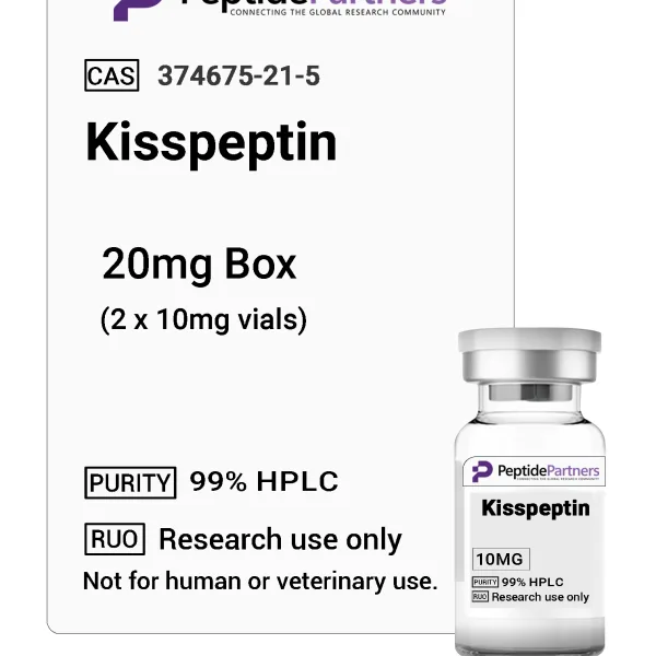
In modern peptide science, few regulatory molecules have transitioned across distinct biological disciplines quite like kisspeptin. Originally identified in oncology laboratories as a suppressor of malignant cellular metastasis, the peptide is now widely recognized as a master upstream driver of the mammalian reproductive axis and an integral node in metabolic signaling. Classified under metabolic and neuroendocrine categories in research catalogs, kisspeptin has become a major target of preclinical investigation into how cellular energy availability dictates central hormonal cascades.
For scientific investigators evaluating kisspeptin for in vitro assays or animal model studies, understanding its structural biochemistry, receptor pathways, and direct links to metabolic physiology is fundamental. This article outlines the scientific discovery of kisspeptin, examines the physiological systems it engages, and provides guidance on selecting analytical-grade reagents and documentation when sourcing from Peptides Partners.
Discovery History: From Metastasis Suppression to Neuroendocrine Architecture
The gene responsible for producing kisspeptin, designated KISS1, was discovered in 1996 by researchers at the Pennsylvania State University College of Medicine. Initially characterized in human melanoma cell lines, the gene product demonstrated a notable capacity to prevent metastasis without inhibiting primary tumor growth, leading to its early designation as “metastin.”
The turning point for endocrinological research arrived in 2003, when multiple independent research groups discovered that loss-of-function mutations in an orphan G-protein coupled receptor, known as GPR54 (now classified as KISS1R), resulted in idiopathic hypogonadotropic hypogonadism in both human pedigrees and murine knockouts. Scientists quickly established that kisspeptin acts as the endogenous, high-affinity ligand for this receptor. Subsequent investigations demonstrated that kisspeptin-expressing neurons located in the hypothalamic arcuate nucleus (ARC) and anteroventral periventricular nucleus (AVPV) form dense synaptic connections with Gonadotropin-Releasing Hormone (GnRH) neurons, effectively positioning kisspeptin at the apex of the hypothalamic-pituitary-gonadal (HPG) axis.
Biochemical Processing and Receptor Signaling Dynamics
The primary transcript of the KISS1 gene encodes a 145-amino-acid precursor protein. Enzymatic cleavage by furin-like endoproteases yields several biologically active isoforms that share a common, highly conserved C-terminal decapeptide sequence. The major forms analyzed in laboratory settings include:
- Kisspeptin-54: The full-length circulating isoform, consisting of 54 amino acids, widely utilized in systems-level endocrine research.
- Kisspeptin-14 and Kisspeptin-13: Intermediate cleavage fragments observed during metabolic degradation cascades.
- Kisspeptin-10: The minimum functional pharmacophore, comprising residues 112–121 of the precursor. It terminates with an amidated RF-amide motif (Arg-Phe-NH2) necessary for receptor binding and activation.
Upon binding to KISS1R, kisspeptin triggers a canonical Gαq/11 signaling cascade. This coupling initiates phospholipase C (PLC) activation, producing inositol 1,4,5-trisphosphate (IP3) and diacylglycerol (DAG). The subsequent mobilization of intracellular calcium stores alongside protein kinase C (PKC) activation depolarizes target neurons. In hypothalamic paradigms, this sustained influx of calcium stimulates the coordinated, pulsatile release of GnRH into the hypophyseal portal bloodstream, sequentially driving the secretion of luteinizing hormone (LH) and follicle-stimulating hormone (FSH).
The Metabolic Axis: Energy Sensing and Neuroendocrine Cross-Talk
While early kisspeptin research focused strictly on pubertal onset and reproductive biology, researchers have increasingly investigated the peptide within the context of metabolic homeostasis. Maintaining an active reproductive axis requires significant bioenergetic resources, and mammalian systems have evolved precise feedback loops to suppress non-essential endocrine programs during periods of starvation, systemic stress, or metabolic imbalance.
Hypothalamic Kiss1 neurons function as direct sensors of peripheral energy status. Published research in preclinical models highlights several key interactions:
- Leptin Signaling: Adipose-derived leptin conveys total systemic energy storage to the brain. While GnRH neurons themselves largely lack functional leptin receptors (LepR), adjacent Kiss1 neurons in the arcuate nucleus express LepR, allowing leptin to upregulate Kiss1 transcription and maintain downstream endocrine tone.
- Orexigenic and Anorexigenic Inputs: Signaling molecules such as ghrelin, neuropeptide Y (NPY), and pro-opiomelanocortin (POMC) engage in complex, direct synaptic communication with kisspeptin networks, modulating neuronal firing rates based on acute nutrient availability.
- Glucose Homeostasis and Adiposity: Preclinical studies utilizing global and tissue-specific Kiss1R knockout rodents have identified phenotypes characterized by impaired glucose tolerance, altered energy expenditure, and increased adiposity, leading investigators to examine the direct metabolic roles of kisspeptin receptors outside the brain, including within pancreatic islet cells and brown adipose tissue.
Core Areas of Preclinical Investigation
In academic and institutional laboratories, kisspeptin is regularly studied across several methodological frameworks:
1. Pulsatile Secretion and Feedback Sensitivity
Investigators utilize kisspeptin analogs to probe the sensitivity of pituitary and hypothalamic tissue to continuous versus intermittent stimulation. These models aid in mapping how steroid feedback mechanisms (such as estrogen positive and negative feedback) alter kisspeptin transcription across distinct hypothalamic nuclei.
2. Metabolic Stress and Reproductive Suppression
Studies evaluating acute fasting, chronic caloric restriction, or diet-induced obesity frequently measure hypothalamic Kiss1 mRNA levels to correlate peripheral metabolic biomarkers with central reproductive suppression or dysregulation.
3. Peripheral Receptor Function
Researchers increasingly examine the expression and biological impact of KISS1R in peripheral tissues, including cardiovascular endothelium, hepatic parenchyma, and placental trophoblasts, highlighting biological mechanisms that extend beyond classical central nervous system signaling.
Methodological Protocols: Sourcing and Analytical Evaluation
Ensuring experimental reproducibility when working with short-chain or full-length regulatory peptides requires rigorous verification of material integrity. Peptide quality can vary significantly depending on synthesis methodologies, cleavage conditions, and post-synthesis handling.
When selecting kisspeptin for research-use-only applications from Peptides Partners, investigators should evaluate laboratory documentation based on clear analytical criteria:
| Analytical Metric | Primary Methodology | Experimental Relevance |
|---|---|---|
| Chemical Identity | Mass Spectrometry (ESI-MS / MALDI-TOF) | Confirms theoretical molecular mass, ensuring the correct amino acid sequence and complete C-terminal amidation. |
| Purity Profile | High-Performance Liquid Chromatography (RP-HPLC) | Quantifies the percentage of target peptide relative to truncated, deleted, or incomplete synthetic byproducts. |
| Physical State | Lyophilization | Ensures compound stability during shipment; reduces moisture content to prevent spontaneous hydrolysis. |
| Reconstitution Parameters | Solubility Profiling | Determines proper sterile vehicle selection (e.g., bacteriostatic water, sterile saline, or specific buffers) for accurate molar dilutions. |
Researchers should also determine the appropriate vial pack configuration based on experimental timelines. Because reconstituted peptide solutions exhibit finite stability—even when stored at -20°C or -80°C—procuring multi-vial packs of lower mass quantities (e.g., individual 2 mg or 5 mg vials) is generally preferable to single high-mass containers. This approach limits freeze-thaw degradation cycles and ensures baseline consistency across multiple experimental replicates.
Summary
Kisspeptin represents an essential bridge linking metabolic status to neuroendocrine regulatory pathways. From its origins as a candidate gene for tumor metastasis suppression to its current role in metabolic and endocrine research, it continues to provide valuable insights into systemic communication between the brain and peripheral organs. As preclinical investigations expand into receptor dynamics, metabolic phenotypes, and neurocircuitry, maintaining access to analytically validated, research-grade peptides remains a fundamental requirement for reliable and reproducible scientific discovery.
Where to buy Kisspeptin for research
Peptides Partners lists Kisspeptin (10mg vials) in 4 pack sizes, all in stock, with price per mg shown for each option. Browse more metabolics compounds in the Peptides Partners research peptide shop. All products are for laboratory research use only.